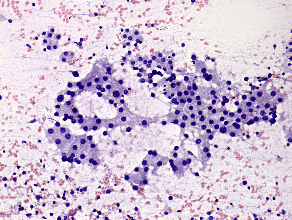
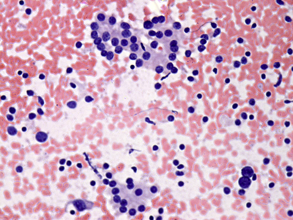
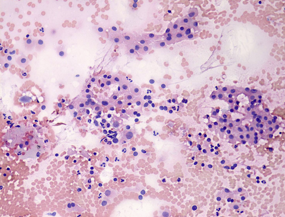
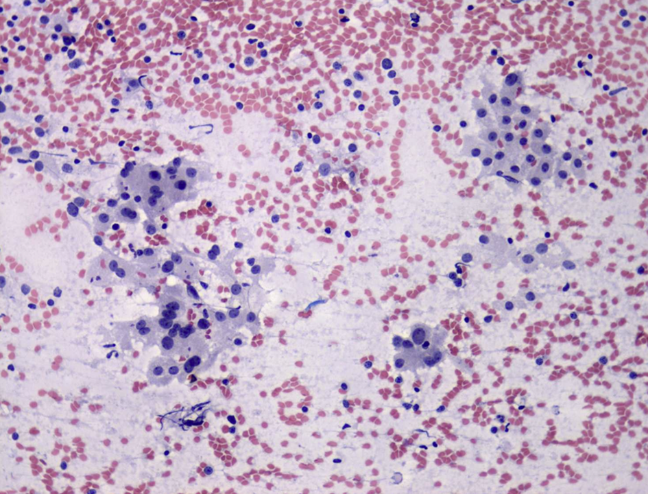
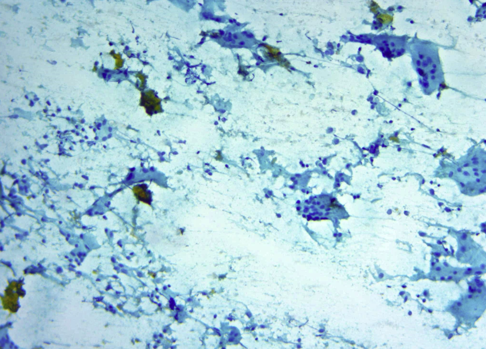
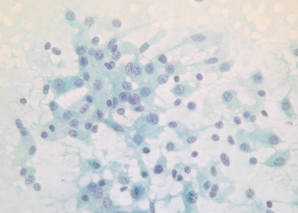
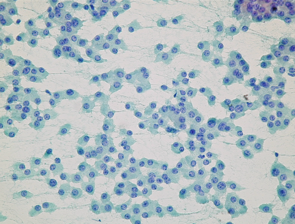
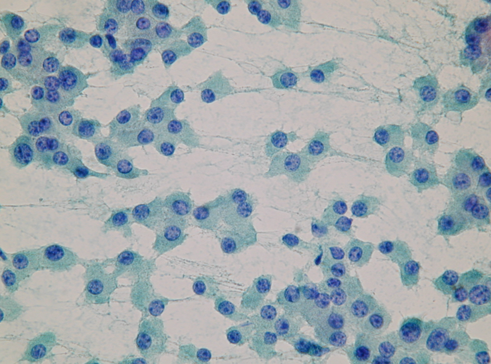
Breast FNA. Be careful with monotonous naked nuclei with nucleoli and admixed lymphocytes in a granular smear background seen in a younger patient. This morphology on FNA is fairly typical of “Lactational Change”. pic.twitter.com/Ta43nxfNgp
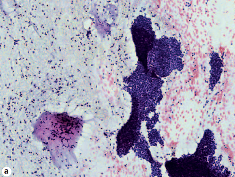
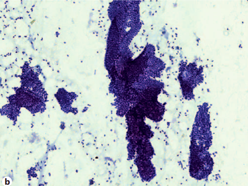
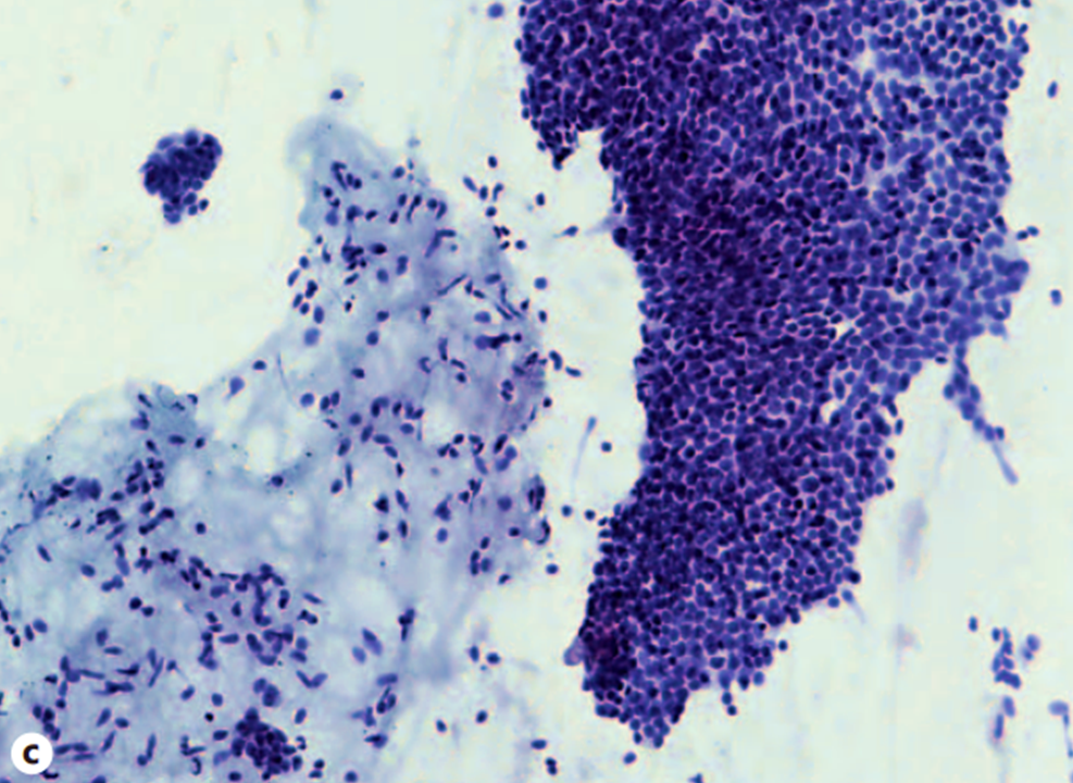
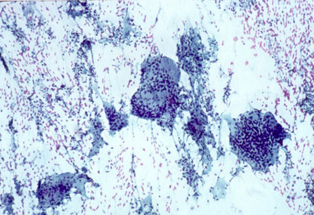
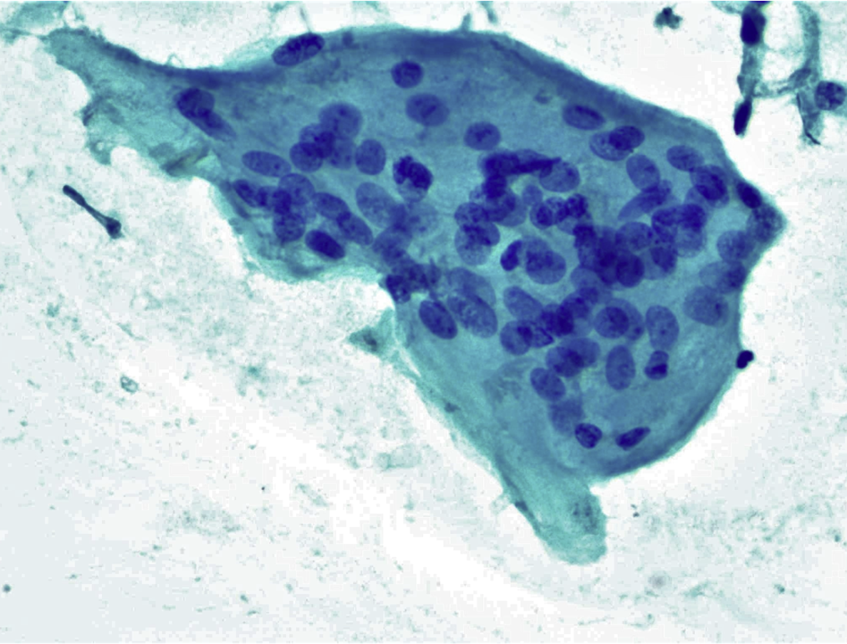
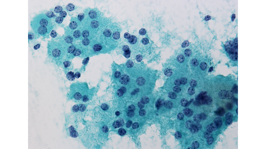
Classic image of a Fibroadenoma. Bening epithelial component with myoepithelial cells. Bipolar and stromal cells in the background. Stromal component fibromyxoid. pic.twitter.com/hzhibE92Vs
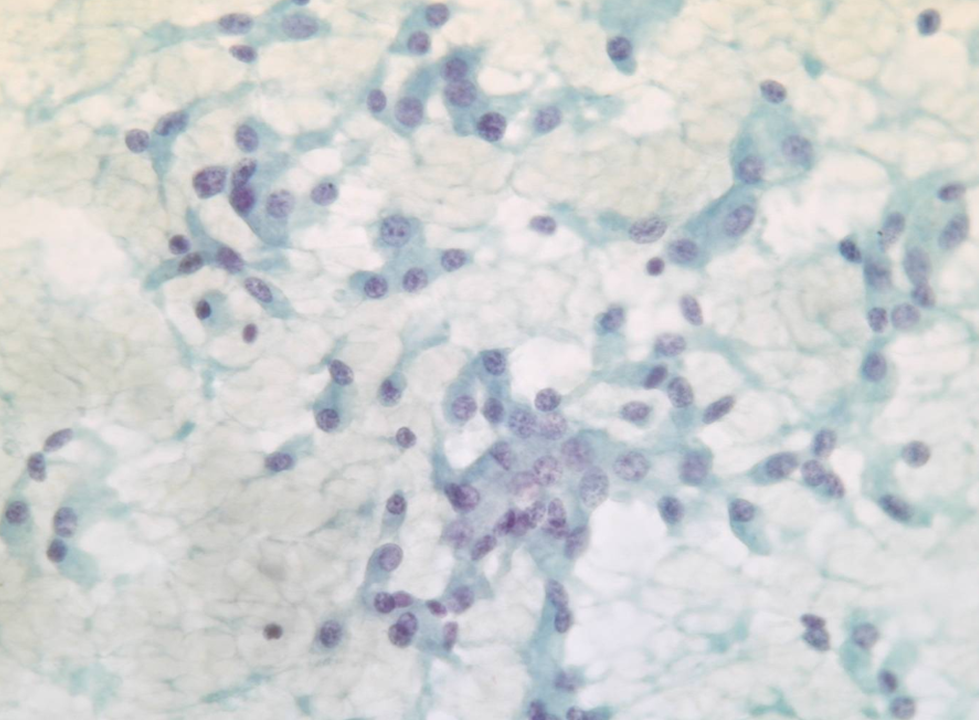
出典:Pinamonti M, Zanconati F: Breast Cytopathology. Assessing the Value of FNAC in the Diagnosis of Breast Lesions. Monogr Clin Cytol. Basel, Karger, 2018, vol 24, pp 58–67 (DOI: 10.1159/000479768) Fibroepithelial Lesions出典:Pinamonti M, Zanconati F: Breast Cytopathology. Assessing the Value of FNAC in the Diagnosis of Breast Lesions. Monogr Clin Cytol. Basel, Karger, 2018, vol 24, pp 58–67 (DOI: 10.1159/000479768) Fibroepithelial Lesions出典:Pinamonti M, Zanconati F: Breast Cytopathology. Assessing the Value of FNAC in the Diagnosis of Breast Lesions. Monogr Clin Cytol. Basel, Karger, 2018, vol 24, pp 58–67 (DOI: 10.1159/000479768) Fibroepithelial Lesions